

Division of Abdominal Imaging
The Division of Abdominal Imaging is a high-volume, research-oriented department specializing in the full spectrum of diagnostic modalities. By integrating Ultrasound, CT, MRI, and Fluoroscopy, the division provides comprehensive diagnostic support backed by the latest clinical research and academic rigor.
Academic Excellence & Innovation
The division is a recognized leader in radiological research, bridging the gap between benchtop science and bedside care.
Specialized Research
Specialized Research
With over 10 publications specifically focused on Dual-Energy CT (DECT), the faculty contributes significantly to the global understanding of advanced imaging.
Future-Ready Programs
Future-Ready Programs:
Multiple ongoing research programs are currently exploring the integration of Artificial Intelligence (AI) to automate detection and improve the accuracy of abdominal diagnostics.
Advanced Ultrasound Capabilities
The ultrasound suite features next-generation technology that goes beyond standard imaging.
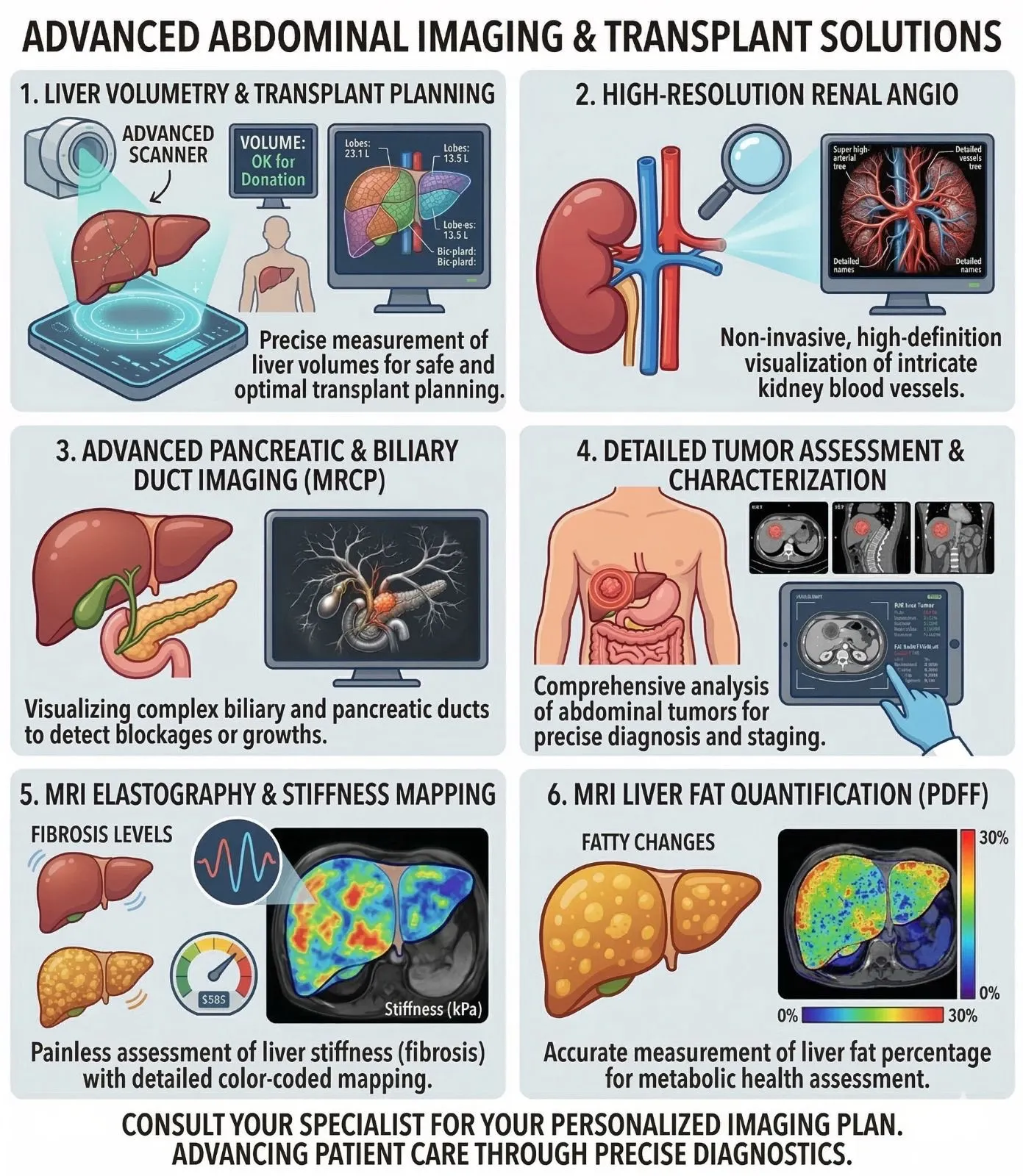
- Tissue Characterization: Capabilities include elastography, viscosity measurements, and fat quantification, providing objective data points for the diagnosis of chronic liver disease.
Ultra-Fast Dual-Source Dual-Energy CT
The facility utilizes the world’s fastest Dual-Source Dual-Energy CT technology, which enables high-speed, motion-free abdominal scans.
- Clinical Impact: This technology allows for high-resolution imaging without the need for patient breath-holding, making it an ideal solution for pediatric, trauma, or elderly patients who may have difficulty remaining still.
Specialized Dual-Energy CT (DECT) Applications
The division leverages DECT to move beyond basic anatomy into material characterization.
Virtual Monoenergetic Imaging
Virtual Monoenergetic Imaging:
- Enhances contrast and helps to identify the lesions which are difficult to visualize in the routine CT.
- Reduces artifacts from metal implants or dense bone.
Renal Stone Characterization
Future-Ready Programs:
Non-invasively identifies the chemical composition of kidney stones, allowing urologists to tailor treatment plans without surgical intervention.
State-of-the-Art 3T MRI
Utilizing high-field 3T MRI technology, the division delivers exceptional image clarity and detail.
- Patient-Centric Design: Advanced sequences are designed to provide high-resolution imaging of the abdomen while the patient breathes naturally, eliminating the stress and physical demand of repeated breath-holds.
Advanced Quantitative Investigations
The department offers specialized investigations that quantify organ health non-invasively, often reducing the need for surgical biopsies.
- MR Elastography: Measures tissue stiffness to evaluate liver fibrosis.
- Metabolic Mapping: Provides precise quantification of liver fat and iron concentration.
- Dynamic MRI: Assesses real-time physiological changes and blood flow within abdominal organs.
AI-Integrated Prostate MRI
To enhance diagnostic precision, the division utilizes AI-integrated reporting for Prostate MRI.
- Precision Workflow: Artificial Intelligence assists radiologists in the detection and grading of lesions, ensuring a highly accurate and streamlined diagnostic process for patients and referring physicians.
Comprehensive Liver and Renal Transplant Support
The division provides end-to-end imaging support for liver transplant candidates and recipients.
Pre-Operative Care
Pre-Operative Care:
Includes precise liver volumetry and MRCP (Magnetic Resonance Cholangiopancreatography) for anatomical mapping.
Post-Operative Care
Post-Operative Care:
Ongoing monitoring ensures the long-term viability of the graft and the health of the recipient.
Dedicated Transplant & Intraoperative Imaging
Expertise extends into the surgical environment through specialized ultrasound monitoring.
- Real-time Guidance: Radiologists provide intraoperative ultrasound to assist surgeons during complex procedures.
- Vascular Monitoring: Post-operative ultrasound is utilized to ensure optimal blood flow and early detection of vascular complications.
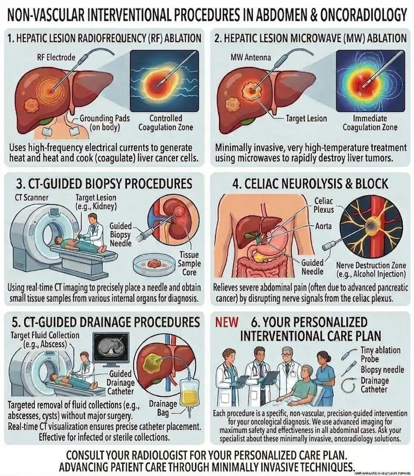
Expert-Led Guided Interventions
The division offers a sophisticated range of non-invasive, image-guided procedures.
Ultrasound-Guided
Ultrasound-Guided:
Specialists perform precise biopsies, Fine Needle Aspirations (FNA), and fluid drainages.
CT-Guided
CT-Guided:
The team provides advanced neurolysis for pain management and targeted ablation of liver tumors, offering a minimally invasive alternative to traditional surgery.
Research & Publications:
Academic excellence with 10+ DECT publications and multiple ongoing research programs including AI.
- Key Paper: "Accuracy of Evaluation of Fatty Liver with Third-Generation Unenhanced Dual-Energy CT and MRI."
- Key Paper: "Dual-Energy Computed Tomography Applications in the Abdomen."
