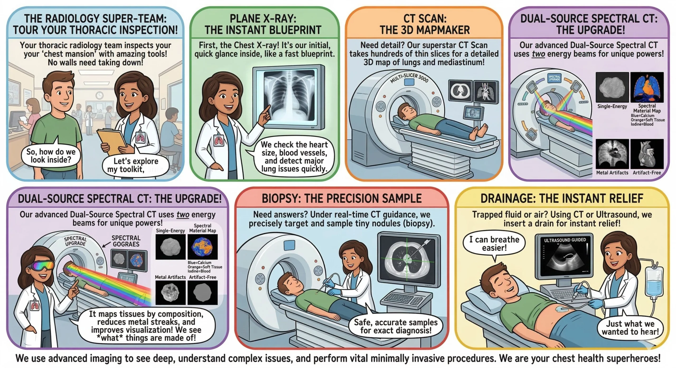
Division of Thoracic Imaging specializes in the imaging, diagnosis, and evaluation of diseases affecting the chest, including the lungs, airways, pleura, mediastinum, diaphragm, and chest wall. Using advanced imaging technologies such as chest X-rays, computed tomography (CT), magnetic resonance imaging (MRI), and ultrasound, the department plays a vital role in the early detection, accurate diagnosis, and monitoring of thoracic diseases.
Thoracic radiology is essential in the assessment and management of various conditions such as lung infections, tuberculosis, lung cancer, interstitial lung diseases, pulmonary embolism, pleural disorders, and mediastinal abnormalities. Thoracic Radiology department closely work with pulmonologists, thoracic surgeons, oncologists, and other healthcare professionals to provide comprehensive patient care.
The department is equipped with modern imaging systems and follows standardized protocols to ensure high-quality diagnostic imaging while maintaining patient safety and radiation protection. Advanced techniques such as high-resolution CT (HRCT), CT pulmonary angiography, Dual energy CT (DECT) and image-guided procedures further enhance the diagnostic capabilities of the department.
Through accurate imaging interpretation, technological innovation, and multidisciplinary collaboration, the Thoracic imaging division contributes significantly to improving patient outcomes and supporting clinical decision-making in thoracic diseases.
The Thoracic Imaging Division collaborates with the Massachusetts General Hospital, Boston, in several research activities and panel discussions. In addition, the division is involved in conducting a multicenter lung cancer screening program.
Research & Publications:
Non-aortic vascular findings on chest CT angiogram: including arch vessels and bronchial arteries.
Long-term follow-up of COVID-19 patients to assess risk factors for the development of post-COVID pulmonary fibrosis
Role of Virtual Monoenergetic Imaging in the assessment of vessel enhancement at segmental levels in third-generation dual-source dual-energy CT pulmonary angiography
Role of ultra-low-dose CT in the detection of pulmonary pathologies
Comparison of Ultrashort TE Lung MRI and HRCT for detection of pulmonary nodules in oncology patients